A Guide to Gel Electrophoresis and Gel Documentation Systems
Amanda2024-08-16T16:28:30+00:00Imagers or Gel documentation systems have become indispensable in molecular biology for capturing and analyzing nucleic acid. Gel Docs and Western blot imagers provide a reliable method for capturing and analyzing gel electrophoresis results, allowing scientists to visualize nucleic acids or proteins. In this post, we’ll explore how these instruments came to be, the various types that are on the market, and their role in modern research.
A Brief History of Electrophoresis
The evolution of gel documentation systems is closely tied to the advancements in molecular biology techniques. In 1807, scientists discovered that particles would separate when a charge was applied. The process, a form of chromatography, can be used for particles and separate compounds. By 1942, Electrophoresis was becoming more common in scientific research, and by the 1960s, it was being applied to the separation of nucleic acids. Until then, scientists relied on ultracentrifugation to separate DNA and RNA, which was time-consuming and required many samples. Attention turned to how DNA might behave when an electrical field was applied. When it was successful, the process was termed Electrophoresis.
Soon, advancements were made, such as swapping UV excitable Ethidium Bromide from radioactive tags and developing higher-definition gel mediums. Imaging and documentation systems were introduced to help more precisely analyze the bands created during Electrophoresis and capture the bands for further analysis. These systems embraced digital imaging technology and have become increasingly sophisticated, adding near-infrared imagining and working with multiple dyes and methods.
Anatomy of Gel Documentation Systems, Imagers, and Scanners in the Lab
In the laboratory, gel documentation systems are primarily used to visualize stained or labeled DNA, RNA, and protein bands separated by gel electrophoresis. After running a gel, it is stained with a fluorescent dye, such as ethidium bromide or SYBR Green, which was introduced in 1999. Other stains include SYBR gold, Texas Red, Deep Purple, Sypro orange, and more, each highlighting a different capture aspect.
The stain binds to the molecules of interest, and the amount that binds is related to the molecular weight; the heavier the molecule, the more dye will bind. The gel is then placed in the documentation system and exposed to UV or visible light.
A UV Transilluminator is the most basic visualization method of an electrophoresis gel. These simple platforms backlight a gel with ultraviolet light, sometimes with selectable wavelengths ranging from 200-850nm, that excite the fluorescent dyes in gels, most often Ethidium Bromide. They are simple and cost-effective ways to visualize the results but require careful handling due to the potential hazards of UV exposure. Safer alternatives include white light and LED lights, which can be used with safer dyes excited by blue light, such as SYBR Safe.
When it is time to document the gel, blocking out ambient light with a darkroom or hood becomes necessary. These hoods block the light and provide a brighter capture and more defined images while protecting the user from UV rays. These hoods range from a small tent over a transilluminator plate to a large cabinet with multiple excitation sources and detectors. Scanner-type imagers like the LiCor systems also block ambient light with a platform door cutting down on the bulk on the bench.
Image capture is done with a CCD camera, which captures an image of the gel, which can be analyzed using specialized software. The higher the resolution, the more information users have to work with. These systems often have auto-focus and auto-exposure for optimized captures. This process allows researchers to determine the size and quantity of the molecules, assess the success of PCR reactions, and verify the results of cloning experiments.
Types of Gel Documentation Systems
Several gel and western blot documentation systems are available, from basic visualization to multiplexing multiple dyes to high-definition cameras and numerous light sources, each designed to meet different laboratory needs and degrees of precision.
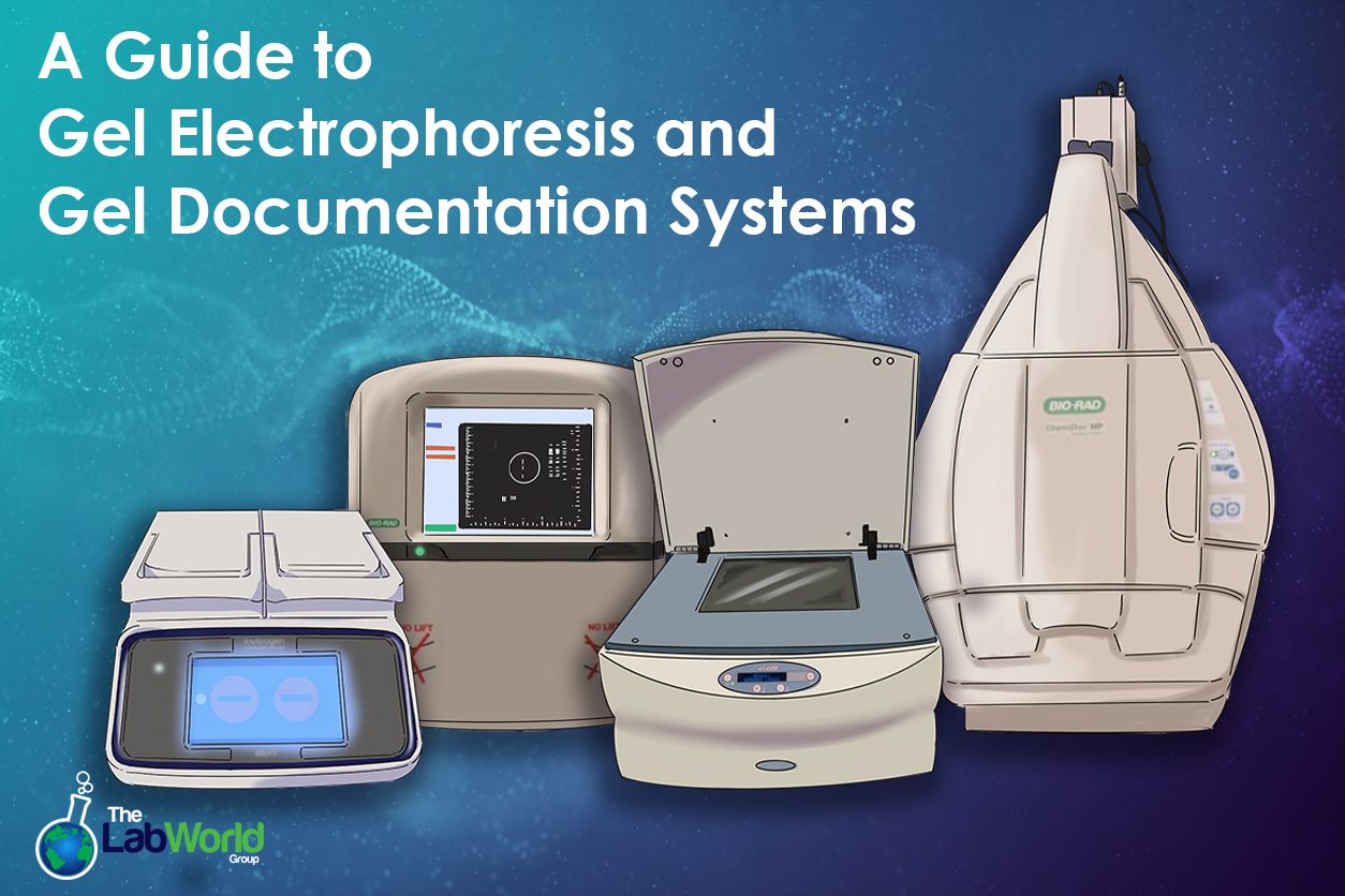
A Chemiluminescence Gel Doc System is popular for working with western blotting techniques or gels but only provides chemiluminescence detection. These systems are highly sensitive and do one thing very well, even working with stain-free techniques and silver stains. If more detection options are needed, look for an imager that can be upgraded to include fluorescent detection, such as the Biorad Chemidoc system. A system with color detection may also include NIR and Far Red as part of the spectrum.
An infrared imaging system is ideal for quantitative analysis and wide dynamic ranges. The CLx from Licor features two detection lasers at 700 and 800nm, with minimal leakage and high signal-to-noise ratios that give you highly precise bands. These scanners also can detect proteins, image small animals and tissue sections, and microwell ELISA.
Multiplexing systems are built to detect and capture multiple targets in the same run. These systems tend to be the most flexible option but are also costly. Detection methods can include luminescent, colorimetric, and radioisotopic signals.
A Phosphorimager works like an x-ray machine where a screen is exposed to a radioactive sample and then scanned with a laser to capture the visible light version. This method is popular for applications like enzyme assays, in vivo imaging, and electrophoretic mobility shift assays.
Gel Documentation and Imager Manufacturers
Several manufacturers dominate the market for gel documentation systems, each offering a range of products tailored to different research needs. Here are a few of the leading companies producing instruments today.
Bio-Rad Gel Documentation
Bio-Rad is known for its GelDoc EZ and ChemiDoc systems. These high-quality, user-friendly documentation solutions are often upgradeable. Their systems are also compatible with robust documentation software that allows users, among other things, to store the images for further analysis, either built into the system or through a controlling PC.
Thermo Scientific Gel Documentation Systems
Thermo Fisher Scientific iBright Imaging Systems are sleek and compact. The FL1500 system multiplexes up to four proteins in a blot and uses a highly sensitive 9.1mp camera. These systems also work with a Green LED transilluminator that excites ethidium bromide while avoiding UV.
Analytik Jena/UVP Imaging Systems
Systems like the Biospectrum Imaging System and the small-scale photo-doc-it systems combine reliability with affordability. The larger hood models can be customized by swapping filters and camera types. On the other end of the spectrum is the compact photo-doc-it system, a perfect entry-level instrument, making its systems popular in academic and research institutions.
Azure Biosystems
Azure Biosystems offers a range of imaging systems, some of which can be upgraded as their use expands. Their 600 Imager, for example, is an exceptionally flexible instrument with a 9-megapixel camera, NIR, RGB, Chemi, UV, blue light, trans white, and true color detection, and powerful built-in software to optimize and analyze after.
LiCor Imagers
LiCor has been building flatbed scanner Odyssey Imaging Systems for years. These systems leverage two lasers at NIR and digital image capture instead of film. The large scanning area simultaneously captures multiple blots, mini-blots, slides, and microplates at a high resolution. These scanners are an ideal choice for quantitative Western blot technology.
Other notable manufacturers include Cytiva Amersham ImageQuants, Typhoon Laser platforms, and Protein Simple Wes System, which is capillary-based and can run 24 simultaneously.
Conclusion
Gel documentation systems have transformed how molecular biologists analyze and document their results. From their rudimentary beginnings to the sophisticated systems available today, these tools have significantly enhanced the accuracy and efficiency of gel analysis. With various systems from leading manufacturers, laboratories can choose the best fit for their specific needs, ensuring that gel documentation remains an integral part of their research workflow.
Choosing a Gel Documentation System or Imaging System can be complicated. Knowing what features are necessary and which may be less critical in getting the desired results. There are many combinations of detectors, filters, and excitation sources. Our team is well acquainted with each machine’s features and limitations and wants to connect you with the right instrument for the task. Don’t hesitate to contact us. We’re here to help!


