How Gel Documentation Evolved
Amanda2024-08-20T12:39:31+00:00Electrophoresis, gel documentation, and Western blot imaging systems are pivotal techniques in molecular biology, allowing scientists to separate, visualize, and analyze proteins and nucleic acids. Over the decades, these technologies have evolved significantly, enhancing our ability to understand the complexities of biological systems, optimize PCR protocols, and test antibiotics and vaccines. Here, we’ll examine the history and development of these essential laboratory methods and how they have evolved to meet the lab’s needs.
The History of Electrophoresis
Electrophoresis, a technique for separating molecules based on size and charge, was introduced in the early 1930s by the Swedish biochemist Arne Tiselius. Considered by many to be the father of Electrophoresis, he developed the “Tiselius apparatus,” an early form of Electrophoresis that could separate proteins in free solution. Tiselius has been the research assistant for a biochemist, Thé Svedberg, at Uppsala University in Sweden; Svedberg discovered that cells were made of molecules, and together, they would separate those molecules through ultracentrifugation. This process was time-consuming, often requiring Tiselius to sleep in the centrifuge room overnight to monitor conditions.
The idea of using electrical fields to separate ions in a solution had been demonstrated in the mid-1800s. Using charged ions as an alternative to centrifugation intrigued Svedberg, who handed the idea to his research assistant, Tiselius, to pursue while he continued his work. He discovered the separation results could be photographed when carefully filtered UV light was applied. As the years went on and the interest in his research grew, he refined his process, using narrower capillaries, buffers that better resisted conducting, and higher voltages.
The Nobel Prize Research
Until then, blood serum was moved to one group during centrifugation. However, when using what would become known as Electrophoresis, he found that blood comprises four components. These results were visualized using Schlieren photography, a photography method to capture fluid flow using light rays bent by changes in density. After he was awarded the Nobel Prize for Chemistry in 1948 for this discovery, the method gained widespread use.
Gel electrophoresis
By the 1950s, zone gel electrophoresis was revolutionizing biochemical studies. PAGE, or polyacrylamide gel electrophoresis, moved away from Tiselius’s Movable Boundary Electrophoresis technique. It allowed for the resolution of proteins based on their size by using a cross-linked gel matrix made of acrylamide and bisacrylamide. This gel matrix improved separation and resolution and became the standard.
In 1960, agarose gel electrophoresis became the method for separating nucleic acids, such as DNA and RNA. The gel matrix, derived from seaweed, provided a robust and reproducible medium for separating large biomolecules.
By 1970, the PAGE technique was further developed by introducing SDS, sodium dodecyl-sulfate. SDS-PAGE produced high-resolution separations by denaturing proteins before Electrophoresis, giving them a uniform negative charge and enabling separation based on size. This development, in combination with isoelectric focusing and protein staining, gave way to a resolution high enough for effective gene mapping.
Protein Dyes and Documentation
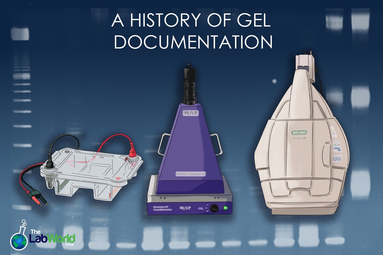
When scientists first started documenting gels, a silver emulsion film was used with instant film. However, the process was cumbersome and lacked sensitivity. Gel documentation systems emerged alongside advancements in Electrophoresis, enabling the capture and analysis of gel images. Around the 1990s, digital imaging systems emerged. High-resolution CCD cameras and software that ensured accuracy and robust analysis streamlined workflows, improved data collection, and fostered reproducible results.
Dyes are essential for visualizing molecules separated by Electrophoresis. Initially, Coomassie Brilliant Blue was the dye of choice for staining proteins in polyacrylamide gels. It binds to proteins, producing a blue color that is easily visible and quantifiable. Ethidium bromide (EtBr) was commonly used for nucleic acids. EtBr intercalates between DNA bases and fluoresces under ultraviolet light, allowing visualization of DNA bands.
Western Blots Imaging emerged in the 1970s, building upon the Southern and Nothern Blot technology of visualizing DNA and RNA fragments, respectively. Western Blot Imagers collect radiographic images of antibody-specific antigens.
Chemiluminescence substrates were introduced in the 1980s, allowing for highly sensitive protein detection on Western blots. This technique involves using an enzyme-conjugated secondary antibody that produces light when exposed to a specific substrate. This technique was further expanded in the 1990s to early 2000s with the introduction of Fluorescent Western Blotting. Introducing fluorescent dyes and secondary antibodies enabled multiplex detection, allowing the researcher to see multiple proteins on the same blot.
The discovery and development of Green Fluorescent Protein (GFP) in the 1990s opened new avenues for live-cell imaging and molecular tracking. GFP and its variants could be genetically fused to proteins of interest, enabling real-time visualization of biological processes. Dyes like fluorescein, rhodamine, and cyanine dyes became popular for labeling biomolecules. These dyes offered high sensitivity and versatility for various applications, including fluorescence microscopy and flow cytometry.
Final Thoughts
The evolution of Electrophoresis, gel documentation, and Western blot imaging systems has significantly advanced our understanding of molecular biology. From the early days of Tiselius’s apparatus to today’s sophisticated fluorescent imaging systems, these technologies have continually improved in sensitivity, specificity, and ease of use. Introducing fluorescent dyes and proteins marked a turning point, enabling more detailed, granular, and dynamic studies of biological systems.
While these instruments play a crucial role in the lab, they can also be cost-prohibitive. One of the best ways to expand your research and streamline your workflow while keeping track of your budget is to buy a vetted, gently used imaging system. We are familiar with these sensitive instruments and have carried out all visualization needs, from simple transilluminators to photo doc systems, chemiluminescent systems, western blots, and more. Let us find the right gel documentation system and technique for the job!


