Anatomy of Gel Documentation Systems and Gel Scanners
Amanda2024-08-22T13:50:30+00:00In scientific research and laboratory diagnostics, the ability to visualize and analyze biological samples such as DNA, RNA, proteins, and cells is crucial. Visualizing DNA bands after gel electrophoresis allows researchers to study genetic mutations or sequence variations.
Gel documentation systems, gel scanners, and laboratory imagers play pivotal roles in this process, offering researchers powerful tools to capture, analyze, and document crucial data. Here, we’ll examine the components essential for these laboratory instruments to understand their functionalities, applications, and technological advancements.
The Basic Components of a Gel Documentation Systems
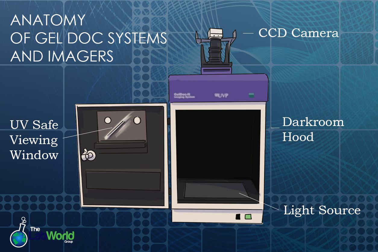
Gel documentation systems are specialized imaging devices used primarily in molecular biology and biochemistry laboratories. They are designed to capture images of biological samples separated on gels through techniques like gel electrophoresis and western blotting. These systems typically comprise a Camera: a CCD or charge-coupled device camera or a CMOS complementary metal oxide semiconductor camera.
A CCD Camera is ideal for low-light situations but yields a high resolution; these cameras have the advantage of a wide dynamic range, spatial resolution, spectral bandwidth, light sensitivity, and capture speed. These digital cameras are more sensitive than your everyday DSLR camera, with less noise and pixel defects and thermoelectric cooling to further reduce noise. A CMOS camera uses photodiodes and consumes less energy (up to 100 times less!) than a CCD camera; they’re less expensive than a CCD camera; however, they are more susceptible to noise and tend to be less sensitive to light.
Gel Documentation Systems also have lenses and/or filter configurations that optimize clarity and contrast and are built to work with specific dyes and more. Depending on the technique, a Gel Doc will have a light source such as UV or White Light. Finally, a hood or a means of blocking ambient room light effectively captures a gel and protects users from UV irradiation. This light-blocking method can look like a large cabinet, a small tent-shaped hood, or the lid of a flatbed scanner.
Specialized Components of a Gel Doc System
Image analysis software is sometimes included with a system depending on how advanced the documentation system is. It may be kept on a separate PC or built into the system. Analysis software provides greater insight, merges different channels into a single image, simplifies a workflow, and can even store images for further analysis. Additional innovations can include touchscreen interfaces and built-in computers that provide user-friendly guidance through each step, making them more accessible.
Beyond white or UV light, gel documentation systems might include multiple colored light options, such as Blue, Blue/Green, and Red LED, that expand the excitation range and avoid the DNA damage that can occur. Systems like the Azure 600 western blot system cover RGB light, chemiluminescence, blue light, UV, and Near Infrared Light. Systems that use EVOS light cubes have an easy-to-use method of quickly switching methods and excitations.
Components of a Gel Scanner
Gel scanners are advanced versions of gel documentation systems, offering enhanced capabilities for automated scanning and digital capture of gel images. They provide high-resolution images suitable for quantitative analysis and archival purposes.
Gel scanners are built differently and don’t have a housing component. Gels are scanned on a platform to capture a high-definition image with high sensitivity. Scanners can capture multiple wavelengths simultaneously. Some, like the LiCor Odyssey, use two solid-state diode lasers, providing excitation at 700 and 800 nm, while the microscope electronics pick out the IR dye from the background fluorescence. After passing to a dichroic mirror, more optics to remove scattered and stray light, and conversion to an electrical signal, a digital signal processor produces an image file that avoids over-saturation and blowout. Scanning surfaces can vary from small to more significant throughput.
Additional Components and Options
Gel documentation systems can be multifunctional beyond Western blotting and Nucleic Acid applications. Gel documentation systems that include microscope integration provide cell and tissue imaging. Imaging software may also offer quantification tools for cell colonies, imaging cellular structures, tissue samples, and protein interactions.
Innovations and looking to the future
Gel documentation systems and Gel Scanners are already versatile tools in the laboratory. Algorithms help to automate processes and recognize patterns. As AI tools grow and learn, these machine-based analyses will pick out patterns the human eye might miss. As technology continues to grow, we can expect more compact systems that take up less space on the bench, use more efficient components, and use less energy.
Conclusion
Gel documentation systems and gel scanners are indispensable tools in modern scientific research. They enable precise analysis and visualization of biological samples. As technology evolves, these instruments will advance our understanding of genetics, molecular biology, and disease mechanisms. When choosing the right tool for your lab, you should consider a few things; budget is undoubtedly one of them. One way to find the most bang for your buck is to buy gently used and thoroughly vetted instruments from a reputable dealer.
One of the lab world group’s most popular departments is Gel Docs. We have carried systems from simple transilluminators to full-spectrum instruments. We’re also familiar with every name-brand maker and how to keep them in top condition. Our inventory changes often, so please don’t hesitate to contact us if you have any questions about a particular Gel Doc or Gel Imaging System.


