A Brief History of Cell Counting
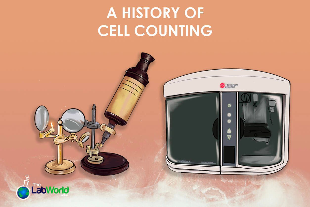
Amanda2024-05-24T13:11:14+00:00Understanding the population of cells or concentration of particles in a fluid can be a useful diagnostic tool. For nearly 300 years, we’ve been observing blood cells since the simple microscope was improved in the mid to late 1600s. While simple microscopes were invented in the late 13th century as an extension of the burgeoning field of eyeglasses, the stacked lenses didn’t have enough magnification power to reveal the tiny universe yet. That was until a Holland inventor named Antoine van Leeuwenhoek found that by rounding the edge of a lens through grinding and polishing, the magnification increased nearly 270x. These compound microscopes opened up this hidden part of our world, and science jumped forward.
In 1665, Robert Hooke used a compound microscope to observe cells and began drawing what he saw. He published a book called Micrographia and pioneered the term “Cell.” When he observed the rectangular structures in a slice of cork, they reminded him of monastery rooms, and he deemed them “cells.” This observation paved the way for cellular theory. Eventually, it would lead to modern cellular theory to include how, within similar species, cells are mostly the same and how energy flows within the cell.
Improving Magnification
Microscopes stayed largely the same for around 200 years after this, with few adopters and fewer innovations. It wasn’t until the 1830s when Joseph Jackson Lister and William Tulley developed a microscope that corrected two significant problems with magnification: Image blurring and chromatic aberration. Once these problems were solved, the microscope made its way into more and more laboratories, and cells became a focus of studies. Adding dyes to slides in the late 1870s by Paul Ehrlich made it possible to see the difference in white cell types. Now, the practice moves beyond observing and describing these structures when it becomes evident that the number of blood cells can vary when a disease is present. The ability to count how many cells are present and to do so accurately becomes more critical.
The First Hemocytometer
In 1874, Louis-Charles Malassez crafted the first hemocytometer, a thick glass slide with a grid and known volume that allowed users to manually count the cell concentration in that volume and use that to calculate the concentration in the overall fluid the sample was taken from. This method is still practiced today; it’s simple, and if your lab lacks a dedicated instrument, you can still get the job done with a hemocytometer slide. However, it’s time-consuming, labor-intensive, and prone to error. To streamline the process and reclaim lost time, engineers and scientists set to work on creating Automated cell counters.
The Coulter Principle
Automated particle and cell counters came around in 1953 thanks to Walter Coulter, who used electric resistance to capture a cell count, known as the Coulter Principle. Naturally resistant to electricity, cells would pass through a single stream between two electrodes conducting a current. When that current is interrupted, a count is recorded. This method also gave way to determine the size of the cell depending on how much resistance was registered as it passed. Soon, the ability to gather other qualitative differences, such as shape and degree of hemoglobinization, allowed hematologists to classify different types of anemia and other afflictions. Blood analysis is among the easiest and least invasive health monitoring methods today thanks to the Coulter Counter.
Rise of Image-Based Analysis
Since those early days of manual counting cells on a slide, flow methods and impedance measurements with the Coulter Principle have been introduced. After this came light scattering and fluorescence techniques to catalog cells individually. One technique that’s become increasingly popular for its simplicity and accuracy is image-based microscopy. Taking principles from manual visualization counting and applying powerful image analysis software with a recognition algorithm significantly increased accessibility to automated cell counting.
Imaged-based cell counting has become more popular over the years due to the more straightforward sample preparation and the overall size of the instrument. Imaged-based automatic cell and particle counters can exist outside a central testing laboratory. However, the drawback of the image-based automatic cell counter is the smaller field of view, so magnification is something to consider when choosing an instrument for a laboratory. As time passes, innovations such as holographic imaging without lenses and imaging chips will increase the power and scope of these helpful diagnostic instruments.
Flow cytometry-based counters provide high acquisition rates and fluorescence sensitivity for speed and high throughput. This cell-counting method allows for a quick analysis of statistically significant populations. However, the tradeoff is a lack of information on sub-structures. As a result, some labs do both and combine the information gathered, which means two separate sample preparations and protocols but a more robust picture.
Final Thoughts
Whether a flow cytometry-based particle counter and high throughput are best for your needs or a deep dive into the cell’s structure, let us help you find the right tool. Our instruments are thoroughly vetted when they come to us and are verified against the original manufacturer’s specifications so you can be sure of accuracy and processing power.
We’ve carried many brands like Thermo Scientific, Invitrogen, and Beckman Coulter and are familiar with their inner workings and software. If you have questions, we have answers. Don’t hesitate to contact us today and let us help you speed up your workflow!


