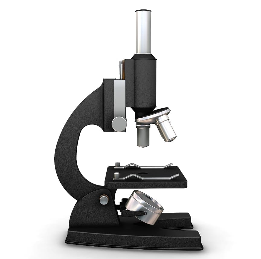
New Nano3 microscope will allow high-resolution look inside cells
The first institution to receive a novel FEI Scios dual-beam microscope is The University of California, San Diego’s Nanofabrication Cleanroom Facility (Nano3). The new microscope will allow research wlong a highly diverse user base, starting from materials science to structural and molecular biology. Nano3 Technical Director Bernd Fruhberger ellaborates: “There is a tremendous interest in utilizing this instrument among faculty from multiple departments. The Departments of Nanoengineering, Materials and Aerospace Engineering, Electrical and Computer Engineering, Chemistry, Physics and Biology at UC San Diego all have projects in need of this tool, and have been actively involved in making the procurement of the tool a reality. The instrument provides state-of-the-art capabilities for cross-sectioning, preparation of sections for Transmission Electron Microscopy and more,” he continues, “but what truly differentiates it is the novel cryo-capability, which will make it possible for cell biologists to see the structures of biological cells in higher resolution to better understand how cells function at a molecular level. This could possibly pave the way for new treatments and drug discovery.”
A new assistant professor in the Department of Chemistry & Biochemistry at the UC San Diego, Elizabeth Villa, and her colleagues at Germany’s Max Planck Institute of Biochemistry, adapted a focused-ion-beam microscope for biological applications during her postdoctoral studies. The Dutch company FEI took to the design into a first-of-a-kind prototype that Villa will develop further in collaboration with the company. UC San Diego has built an academic tradition that is reflected in the work of biochemist Roger Tsien. He won the 2008 Nobel Prize in Chemistry for the discovery and development of the green fluorescent protein. “What I’m doing is similar,” explains Villa, “only I’m using electron microscopy, which gives us higher-resolution images. The idea behind our method is to bring together people who do structural biology with people who do cell biology by using a new tool that will allow us to see the structures of the cells, at high resolution, and better understand what molecules are doing.”


